

The Benefits of Using Ultrasound in Pelvic Floor Physical Therapy
As pelvic floor physical therapists, we rely on a combination of clinical skill, patient feedback, and evidence-based tools to guide assessment and treatment. One tool that’s transforming how we evaluate and train the pelvic floor is real-time ultrasound imaging.
Unlike traditional internal exams—which provide valuable but subjective information—ultrasound offers a visual, dynamic, and objective look at how the pelvic floor and core muscles are functioning in real time.
What Can Ultrasound Add to a Pelvic Floor Assessment?
1. Visual Feedback for Pelvic Floor and Core Activation
Ultrasound allows both the therapist and patient to actually see what’s happening during pelvic floor and core muscle activation, relaxation, and breath coordination.
Patients can visualize a contraction, Valsalva, or relaxation pattern, helping them connect what they feel with what’s actually happening.
This immediate feedback makes training more engaging and effective, often accelerating their progress.
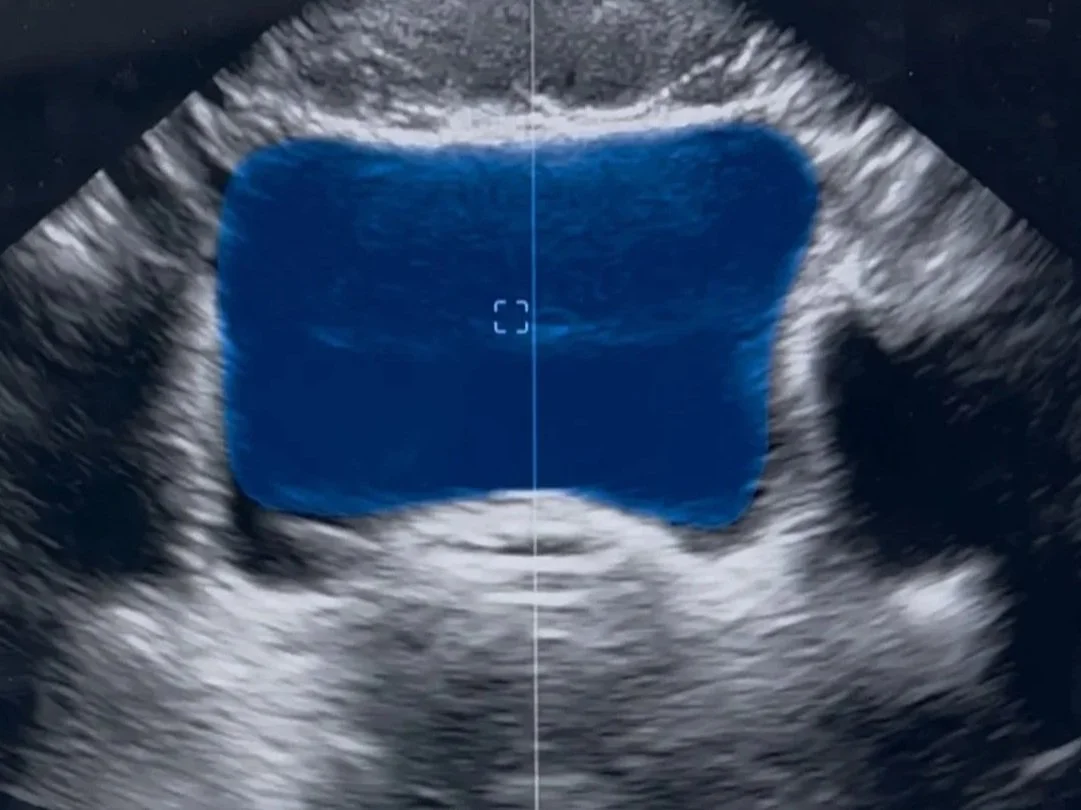
Image: blue represents the amount of urine in the bladder. The bladder is outlined in white. As I contract my pelvic floor I can assess symmetry of contraction and ability to relax after various types of contractions by assessing how the base of the bladder moves in relationship to what the pelvic floor muscles are doing.
2. Dynamic Functional Assessment
Beyond simple contraction tests, ultrasound lets us assess how the deep core and pelvic floor respond during functional movements—like single-leg stance, marches, or straight leg lifts. This dynamic approach provides insight into how these muscles coordinate during movement, helping us identify compensations or timing issues that might otherwise go unnoticed.
3. Pre- and Post-Void Analysis
Using ultrasound, we can visualize bladder filling and emptying patterns. This can reveal whether the bladder is fully emptying or if pelvic floor overactivity might be contributing to urinary symptoms.
4. More Informed and Individualized Treatment Planning
Real-time imaging guides decision-making by giving objective data. Whether the goal is to build strength, improve relaxation, or retrain coordination, ultrasound helps tailor each patient’s pelvic floor training to their specific needs.
How Ultrasound Improves Treatment Outcomes
Increased Efficiency and Precision
When we combine hands-on assessment with imaging, treatment becomes more focused and guided. We can identify exactly how muscles are engaging—so we’re not guessing. This can mean fewer sessions and faster results.
Better Patient Outcomes
Because ultrasound supports both diagnosis and biofeedback, it enhances the accuracy of our clinical reasoning and the effectiveness of our interventions. Research suggests that visual feedback leads to better muscle control and long-term outcomes for patients with pelvic floor dysfunction.
A Non-Invasive and Comfortable Option
For some patients, internal exams can feel intimidating or uncomfortable. Ultrasound provides a non-invasive way to assess pelvic floor function, making the evaluation process more accessible and empowering.
Empowering Patient Education
Perhaps one of the most profound benefits is the educational component. When patients can see their pelvic floor in action, it transforms their understanding of their own body. They become active participants in their care—more aware, more confident, and more connected to their healing process.
The Bottom Line
Integrating ultrasound into pelvic floor physical therapy bridges the gap between subjective assessment and objective data. It helps therapists deliver more precise, evidence-based care—while helping patients feel more informed and empowered in their recovery.
Refrences:
Krasnopolsky N, Ami NB, Dar G. Ultrasound Assessment and Self-Perception of Pelvic Floor Muscle Function in Women with Stress Urinary Incontinence in Different Positions. Diagnostics (Basel). 2024;14(19):2230. Published 2024 Oct 6. doi:10.3390/diagnostics14192230
Frawley H, Shelly B, Morin M, et al. An International Continence Society (ICS) report on the terminology for pelvic floor muscle assessment. Neurourol Urodyn. 2021;40(5):1217-1260. doi:10.1002/nau.24658
Smith M, Donnelly GM, Berry L, Innes S, Dixon J. Point of care ultrasound in pelvic health: scope of practice, education and governance for physiotherapists. Int Urogynecol J. 2022;33(10):2669-2680. doi:10.1007/s00192-022-05200-x
Teyhen DS, Gill NW, Whittaker JL, Henry SM, Hides JA, Hodges P. Rehabilitative ultrasound imaging of the abdominal muscles. J Orthop Sports Phys Ther. 2007;37(8):450-466. doi:10.2519/jospt.2007.2558